Home | Surgeries | Tibial Plateau Leveling Osteotomy (TPLO) Surgery for Dogs
Tibial Plateau Leveling Osteotomy (TPLO) Surgery for Dogs
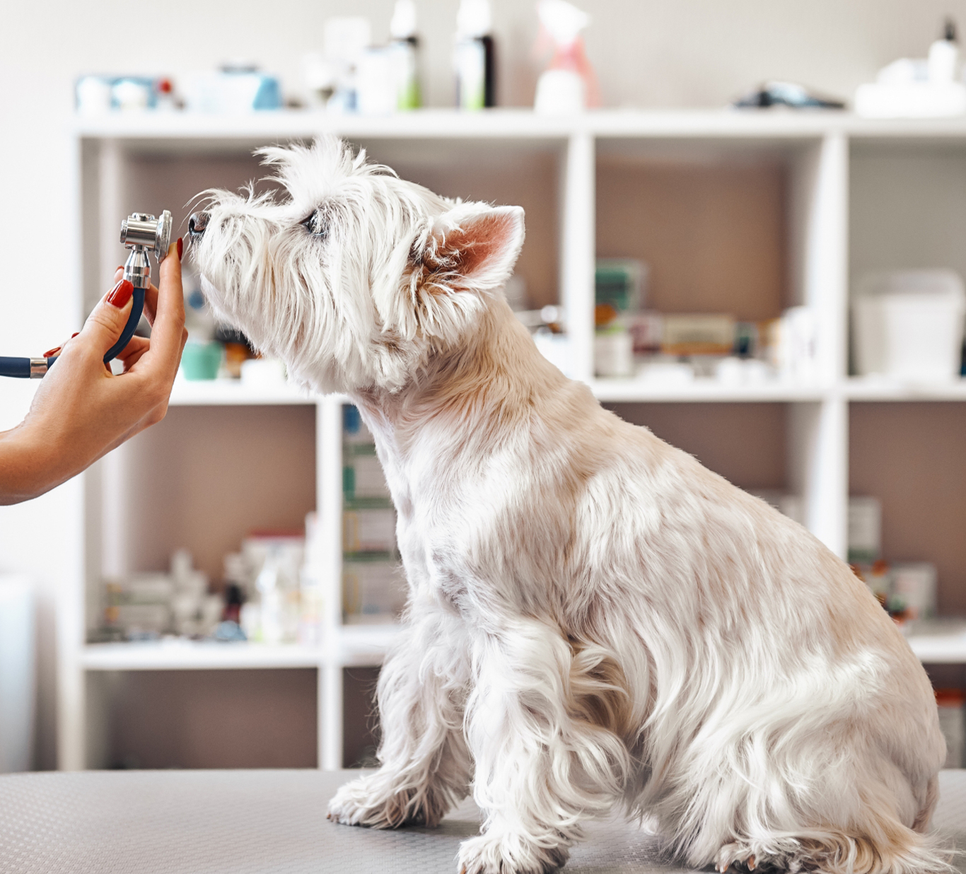
Tibial Plateau Leveling Osteotomy (TPLO) Surgery for Dogs: Cost, Recovery & Success
What is TPLO Surgery for Dogs?
TPLO surgery is the most effective procedure for treating a torn ACL (cranial cruciate ligament) in dogs. This procedure alters the tibial plateau angle to eliminate joint instability, reduce pain, and prevent arthritis. At Canton Animal Hospital, we offer advanced orthopedic surgeries like TPLO to ensure faster recovery and long-term success.
Why choose TPLO surgery?
Best for active, large-breed, and working dogs
Restores knee function & prevents future injuries
Reduces the risk of arthritis & long-term pain
Understanding the Canine Stifle Joint
The canine stifle joint is similar to the human knee but has key anatomical differences. Unlike humans, dogs experience anterior tibial thrust—a forward sliding motion of the tibia during movement. While humans rely on an ACL for knee stability, dogs do not require this ligament if the tibial plateau angle is adjusted to approximately 6 degrees. TPLO surgery modifies the tibial slope to stabilize the joint, allowing dogs to regain function without relying on the CCL.
The Function of the Cranial Cruciate Ligament (CCL) in Dogs
The cranial cruciate ligament (CCL) is a key stabilizing structure in a dog's stifle joint (equivalent to the human knee).
Its primary function is to prevent the femur from sliding backward along the slope of the tibia (shin bone).
This ligament also helps limit excessive internal rotation of the tibia, providing stability when the knee is flexed.
Featured Resources

We Welcome New Patients!
We're always happy to give your furry friend care at our hospital. Get in touch today!
Contact Us

Causes of Cranial Cruciate Ligament (CCL) Injuries in Dogs
CCL injuries in dogs can result from both trauma and degeneration, with many cases involving a combination of both factors. Dogs are particularly prone to CCL tears, often due to underlying ligament degeneration that makes them more susceptible to injury.
Traumatic CCL Injuries
Most commonly occur when a dog suddenly rotates the stifle joint while it is flexed at
20 to 50 degrees.
Can also result from hyperextension of the knee due to forceful impact.
Degenerative CCL Injuries
More common in large-breed dogs as they age.
Can result from conformational abnormalities, such as straight rear limbs, which place extra stress on the knee.
Immune-mediated joint diseases can weaken the ligament over time.
Increased Tibial Plateau Angle (TPA) may contribute to excessive strain on the ligament, leading to gradual failure.
Risk Factors for CCL Injuries in Dogs and Cats
Obesity: Overweight dogs and cats have a significantly higher risk of CCL rupture. Even minor incidents, like stumbling, can cause injury.
Luxating Patella: Dogs with a luxating patella (kneecap dislocation) are more likely to develop a CCL tear, particularly in small and toy breeds.
Bilateral Ruptures: Dogs that rupture one CCL have a 40-50% chance of injuring the other within two years.
Breed Predisposition: Some breeds, including Newfoundland's, Rottweilers, and Labrador Retrievers, are more prone to early-onset bilateral CCL injuries (often between 1 to 2 years of age).


The Impact of a CCL Injury
A CCL rupture or tear is the most common knee injury in dogs. Without treatment, it causes:
Severe pain and lameness
Joint instability, leading to further damage
Osteoarthritis (OA) due to cartilage wear
Early diagnosis and treatment, such as TPLO surgery, can significantly improve mobility and quality of life for affected pets.


Diagnosis of Cranial Cruciate Ligament (CCL) Injuries in Dogs
Accurate diagnosis of CCL injuries is essential for determining the best course of treatment. Dogs of any age or breed can be affected, though young, active, large-breed dogs are the most commonly diagnosed. CCL injuries are rare in cats.
Clinical Presentation of CCL Injuries
CCL injuries can present in three forms:
Acute Injury – Sudden onset of non-weight-bearing or partial-weight-bearing lameness.
Chronic Injury – Prolonged weight-bearing lameness, often worsening over time.
Partial Tears – Mild, intermittent lameness that worsens with activity and improves with rest, making early diagnosis challenging.
Some dogs may also experience bilateral (both knees) CCL ruptures, especially in chronic cases.


Physical Examination Findings
Acute CCL Tears: Dogs with a complete tear may be apprehensive during the examination due to pain. Muscle contraction can make instability harder to detect.
Joint Effusion (swelling): Can often be felt next to the patellar tendon.
"Sit Test": Dogs with CCL injuries often resist fully flexing the knee while sitting.
Periarticular Fibrosis ("Buttress Sign"): Thickening of tissues inside the knee, common in chronic cases.
Cranial Drawer Movement: A key diagnostic test performed with the dog in lateral recumbency. Instability is tested in multiple joint positions (extended, standing angle, and 90-degree flexion). Cranial drawer movement refers to the excessive forward (cranio-caudal) motion of the tibia in relation to the femur due to CCL injury. This instability is one of the primary signs of a torn cruciate ligament and is a key factor in diagnosing the condition.
Tibial Compression Test: Often easier to perform than a cranial drawer test, helping confirm CCL instability. Cranial tibial thrust is defined as cranial movement of the tibial tuberosity in the cranial cruciate–deficient stifle when the hock is flexed and the gastrocnemius muscle contracts.
Understanding Cranial Tibial Thrust in CCL Injuries: Cranial tibial thrust occurs when body weight and muscle forces (such as from the quadriceps and hamstrings) push the proximal tibia forward, creating knee instability.
Normally, the cranial cruciate ligament prevents this motion.
When the CCL is torn, this instability results in cranial drawer movement & positive tibial compression test.
Surgical correction (such as TPLO surgery) aims to reduce the tibial slope to around 5-6.5 degrees, eliminating the excessive cranial tibial thrust while avoiding excessive stress on the caudal cruciate ligament.


Diagnostic Imaging for CCL Injuries
Radiographs (X-rays)
Used to rule out other causes of lameness.
Common findings in chronic or partial CCL tears:
Fat pad compression (“fat pad sign”) and joint capsule swelling due to fluid buildup.
Osteophyte (bone spur) formation on the trochlear ridge, tibial plateau, and patella.
Thickening of the fibrous joint capsule and subchondral sclerosis (increased bone density).
MRI for CCL Evaluation
MRI can provide detailed imaging of the cruciate ligament, especially in unclear cases.
Arthroscopy for Joint Examination
A minimally invasive procedure used to directly visualize the CCL, menisci, and cartilage.
Helps identify ligament tears, fibrillation, and discoloration associated with CCL damage.
Differential Diagnosis of CCL Injuries in Dogs
Several orthopedic conditions can mimic the clinical signs of a cranial cruciate ligament (CCL) injury, making differential diagnosis essential for proper treatment. Common conditions that should be ruled out include:
Mild joint sprains or muscle strains
Patellar luxation (dislocation of the kneecap)
Caudal cruciate ligament injury (less common than CCL tears)
Primary meniscal injury (without ligament damage)
Long digital extensor tendon avulsion
Primary or secondary arthritis
Immune-mediated arthritis
Partial Rupture of the Cranial Cruciate Ligament
A significant number of stifle lameness cases are caused by partial CCL ruptures, which can be more difficult to diagnose than complete tears. Labrador Retrievers and Rottweilers are particularly prone to partial CCL tears at a young age (6-24 months), often affecting both knees and mimicking hip dysplasia. However, CCL issues typically cause more severe lameness than hip dysplasia and should be treated first.
Clinical Signs of a Partial CCL Tear
Similar to complete ruptures but less severe.
Lameness worsens with activity but improves with rest.
Secondary arthritis develops more slowly compared to full ligament ruptures.
Effusion (joint swelling) may be present, indicated by the fat pad sign on X-rays.
Understanding CCL Structure and Injury Patterns
The cranial cruciate ligament (CCL) consists of two key bands:
Craniomedial Band (CrMB) – Taut in both flexion and extension.
Caudolateral Band (CdLB) – Taut only in extension.
How the Type of Injury Affects Diagnosis
Hyperextension injuries often damage the CdLB, leaving the CrMB intact, resulting in no detectable drawer motion.
Drawer motion in flexion (CdLB is relaxed).
No drawer motion in extension (CdLB remains taut).
Twisting or rotational injuries typically affect the CrMB, leading to pain during full extension of a stable but swollen stifle is highly suggestive of a partial CCL tear.
Diagnosing Partial CCL Tears
Partial tears are often overlooked due to minimal drawer motion. However, they should always be considered in medium to large-breed dogs experiencing stifle pain.
Radiographs (X-rays) – The fat pad sign and presence of osteophytes (bone spurs) indicate possible CCL damage.
MRI, Arthroscopy, or Ultrasound – Advanced imaging can confirm partial tears before surgery.
Surgical Exploration – If physical findings are inconclusive, exploratory surgery may be necessary to assess the ligament.
Surgical Approach for Partial CCL Tears
Surgeons should treat partial ligament tears the same way as complete ruptures, as the ligament loses its functionality once damaged. If osteoarthritis is present without an obvious cause (e.g., patellar luxation, meniscal damage, or joint disease), a CCL tear should be suspected.
By using advanced imaging techniques and careful ligament evaluation, early intervention can prevent long-term joint damage and improve treatment outcomes for affected dogs. Learn more about partial CCL tear.
Tibial Osteotomy Techniques for torn CCL in Dogs
Tibial osteotomy procedures, such as Tibial Plateau Leveling Osteotomy (TPLO) and Tibial Tuberosity Advancement (TTA), offer an innovative approach to treating cranial cruciate ligament (CCL) ruptures in dogs. Instead of reconstructing the damaged ligament, these surgical techniques alter the geometry of the stifle joint, effectively eliminating cranial tibial thrust—a key factor in joint instability during movement.
How Tibial Osteotomy Works
Unlike traditional ligament repair methods, TPLO and TTA work by reshaping the tibia, reducing the shear forces between the femur and tibia. Learn more about TTA.
These procedures don't eliminate cranial drawer movement, which is the forward motion of the tibia relative to the femur, detected during orthopedic examinations.
Immediately after surgery, cranial drawer movement may still be present, but cranial tibial thrust should be eliminated both at rest and during movement.
Over time, capsular fibrosis (the natural thickening of joint tissue) helps reduce static cranial drawer motion, further stabilizing the joint.
Tibial Plateau Leveling Osteotomy (TPLO) Surgery in Dogs
Tibial Plateau Leveling Osteotomy (TPLO) is a highly effective surgical procedure developed by Dr. Barclay Slocum to treat cranial cruciate ligament (CCL) injuries in dogs. Dr. Slocum identified the concept of anterior tibial thrust, where the tibia naturally moves forward during weight-bearing. Normally, the CCL stabilizes the knee, much like a rope holding a wagon on a hill. However, when the ligament is torn, this instability causes lameness, pain, and osteoarthritis.
Instead of attempting to reconstruct the CCL, which has shown limited long-term success, TPLO surgery modifies the tibial plateau angle to eliminate instability and restore proper biomechanics to the stifle joint.


How TPLO Surgery Works
The proximal tibia (top of the shinbone) is carefully cut and rotated to change the slope of the tibial plateau.
A specialized bone plate is used to stabilize the osteotomy, ensuring proper healing.
By reducing the tibial plateau angle, the cranial tibial thrust is neutralized, allowing the dog to bear weight without abnormal joint movement.
TPLO is a technically advanced procedure with a steep learning curve.
Complications can be severe if the procedure is not performed correctly.
Experienced orthopedic surgeons are recommended for optimal outcomes.
Meniscal Injuries and TPLO Surgery
Inside the knee joint are menisci, specialized cartilage structures that act as shock absorbers between the femur and tibia. When the CCL ruptures, the medial meniscus is frequently damaged:
Acute Meniscal Tears – Occur at the time of injury.
Chronic Meniscal Damage – Due to prolonged joint instability, leading to crushing and shredding of the caudal horn of the medial meniscus.
Because of the high risk of meniscal damage, TPLO surgery often includes meniscal repair or removal to prevent further joint deterioration.


TPLO Surgery Procedure for Cranial Cruciate Ligament Repair in Dogs
Tibial Plateau Leveling Osteotomy (TPLO) is a precise orthopedic procedure that requires detailed preoperative planning to achieve the best results. Before surgery, radiographs (X-rays) are taken to measure the dog's tibial plateau angle (TPA) and calculate the necessary rotation to achieve an optimal final angle of 5 to 6.5 degrees.
Step-by-Step TPLO Surgical Procedure
1. Stifle Joint Exploration
The stifle joint is examined via either medial arthrotomy or arthroscopy.
If meniscal damage is present, it is repaired or removed to prevent further joint deterioration.
2. Accessing the Proximal Tibia
A medial incision is made to expose the proximal tibia.
Muscle insertions (gracilis, semitendinosus, caudal belly of sartorius) are lifted off the bone, while preserving the medial collateral ligament.
3. Performing the Osteotomy (Bone Cut)
A TPLO jig is applied to guide a curved osteotomy (bone cut) on the medial tibia.
Using a biradial saw blade, the proximal tibia fragment is rotated to the new TPA angle, calculated based on the patient’s individual measurements.
Bone fragments are compressed and secured using a TPLO plate and screws, made of
surgical-grade stainless steel.


TPLO Implants and Fixation
Different types of bone plates and screws are used to stabilize the tibia post-surgery. At our hospital, we use the TPLO Locking System (Plate and Screws) from DePuy Synthes Vet, which ensures maximum strength and stability during healing.
TPLO vs. Other ACL Surgery Options
Best Functional Outcomes – TPLO is widely regarded as the gold standard for CCL repair, allowing even working and performance dogs to return to high activity levels.
Slower Progression of Arthritis – Studies suggest that arthritis progression is slower in dogs that undergo TPLO compared to MRIT (Modified Retinacular Imbrication Technique). However, some degree of arthritis will develop over time regardless of surgical method.
By eliminating cranial tibial thrust, TPLO surgery restores stability, reduces pain, and significantly improves long-term mobility in dogs suffering from CCL injuries.
TPLO Surgery for Small Dogs
TPLO surgery is highly effective for small and toy breeds, especially those with steep tibial slopes that increase ACL injury risk
Post-Operative Care and Potential Complications After TPLO Surgery
While Tibial Plateau Leveling Osteotomy (TPLO) is a highly effective treatment for cranial cruciate ligament (CCL) injuries, proper post-operative care is crucial for a successful recovery.
Potential Post-Surgical Complications
Although complications are rare, some potential risks include:
Delayed or abnormal bone healing
Loosening or breaking of the TPLO plate and screws
Post-surgical infections
How to Minimize Complications During Recovery
To ensure proper healing, we recommend:
Strict crate rest – TPLO patients should be confined to a crate or small space to prevent excessive movement.
Regular X-rays (radiographs) – Follow-up X-rays at 4- and 8-weeks help assess healing progress.
Gradual rehabilitation exercises – Supervised home exercises aid in recovery, with options for continued care at an Animal Rehabilitation Center.
Full healing may take up to 16 weeks in some dogs, requiring patience and careful monitoring.
TPLO Surgery Recovery & Aftercare
Most dogs recover in 8 to 12 weeks with proper post-op care.
Expected Recovery Timeline After TPLO Surgery
3 to 7 Days Post-Surgery – Most dogs will begin partially bearing weight on the operated leg.
3 to 4 Weeks Post-Surgery – Dogs typically walk comfortably with only a slight limp.
8 to 16 Weeks Post-Surgery – The bone heals completely, and most dogs regain near-normal function with proper post-op care.
Activity must be restricted during this period to avoid complications.
We provide detailed post-operative care & rehabilitation plans to ensure a smooth recovery.
2 weeks after bilateral TPLO surgery
24 hours after bilateral TPLO surgery
TPLO Surgery Success Rate & Long-Term Prognosis
How Successful Is TPLO Surgery?
TPLO has a 90-95% success rate, restoring full mobility in most dogs.
It eliminates cranial tibial thrust, preventing arthritis.
Most dogs return to their pre-injury activity levels within a few months.
Will My Dog Be Pain-Free After TPLO?
Yes! TPLO significantly reduces pain & inflammation over time.
Dogs regain full weight-bearing on the leg within weeks after surgery.
How Much Does TPLO Surgery Cost?
The cost of TPLO surgery depends on factors like your dog's size, and anesthesia time. On average, TPLO surgery costs between $3,500 and $4,500.
At Canton Animal Hospital, we offer:
Transparent pricing with no hidden fees
Contact us for a Free Consultation & detailed TPLO surgery estimate!
Choosing a Skilled Veterinary Surgeon for TPLO Surgery
TPLO is a technically advanced procedure with a steep learning curve. It is best performed by an experienced orthopedic surgeon with specialized TPLO certification.
Why Choose Dr. Ajaib Dhaliwal for TPLO Surgery?
Dr. Dhaliwal is Slocum TPLO Certified, ensuring advanced training and expertise.
He has extensive experience in orthopedic procedures, trained under:
Dr. Terri Zachos (Former Assistant Professor at MSU) Dr. Claude Gendreau (Board-Certified Orthopedic Surgeon, Veterinary Orthopedics Center)
Free TPLO Consultation – Dr. Dhaliwal offers second opinions and referral consultations
for TPLO surgery.
If your pet is experiencing CCL injury symptoms, schedule a free TPLO consultation with Dr. Dhaliwal today!
At Canton Animal Hospital, we perform a variety of orthopedic surgeries to help pets recover from joint injuries and improve mobility. Each procedure is carefully selected based on the individual needs of the patient to ensure the best possible outcome.
Extracapsular Lateral Suture (MRIT) – Best for small to medium-sized dogs.
Tibial Plateau Leveling Osteotomy (TPLO) – The gold standard for large, active dogs.
Tibial Tuberosity Advancement (TTA) – A less invasive alternative to TPLO.
General Orthopedic Procedures – We offer surgical solutions to support joint and ligament health.
Frequently Asked Questions (FAQs) About TPLO Surgery
At Canton Animal Hospital, we understand that pet owners have many questions about Tibial Plateau Leveling Osteotomy (TPLO) surgery for dogs. Below, we address some of the most frequently asked questions and clear up common misconceptions about TPLO surgery.


Related Articles about Surgeries
Is your pet scheduling surgery soon? Explore our resource center for additional articles that may be of interest.


Featured Resources
We Welcome New Patients!
We're always happy to give your furry friend care at our hospital. Get in touch today!
Contact UsHelpful Links
Additional Information