Home | Surgeries | Cranial Cruciate Ligament Repair: MRIT/LSS Surgery for Dogs
Cranial Cruciate Ligament Repair: MRIT/LSS Surgery for Dogs
Cranial Cruciate Ligament Repair: MRIT/LSS
Cranial Cruciate Ligament (CCL) injuries are among the most common orthopedic conditions in dogs, leading to pain, joint instability, and arthritis. The Modified Retinacular Imbrication Technique (MRIT), also known as Lateral Suture Stabilization (LSS), is a widely used surgical technique to restore knee stability and improve mobility. This page provides a comprehensive guide to understanding CCL disease, its symptoms, diagnosis, surgical treatment options, and postoperative care. If your pet is experiencing lameness or discomfort, timely intervention can significantly improve their quality of life.
What is Anterior Cruciate Ligament Disease?
Anterior cruciate ligament disease is one of the most common orthopedic conditions in dogs, often leading to degenerative joint disease (arthritis) in the knee.
It is primarily caused by a degenerative process rather than trauma or athletic injury, with traumatic CCL ruptures accounting for only 5-10% of cases.
It can affect all dog breeds and sizes but is most common in large breeds.
The ligament may gradually weaken and partially tear over time before completely rupturing during regular activity.
The exact cause remains unknown, but limb conformation and genetics may contribute to its development.
Partial ligament tears can be difficult to diagnose and frequently occur in both legs simultaneously.
When the ligament tears, the stifle joint becomes unstable, causing a "drawer movement" between the femur and tibia, leading to pain, inflammation, and potential damage to the meniscus.
In about 50% of cases, the medial meniscus (inner joint cartilage) is torn and requires partial removal to relieve pain and prevent further joint damage.
Featured Resources

We Welcome New Patients!
We're always happy to give your furry friend care at our hospital. Get in touch today!
Contact Us
Symptoms of CCL Disease
Limping
Holding the hind limb up
Sitting with the leg extended outward
Stiffness, especially after exercise
Pain upon movement or touch
Joint swelling
Occasional clicking sounds when walking

Diagnosis of CCL Disease
A thorough medical history and physical examination, including tests such as the "cranial drawer" and "tibial thrust" tests.
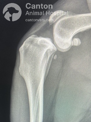
X-rays to evaluate arthritis severity and guide treatment decisions.
Sedation or anesthesia is required for a definitive diagnosis to minimize pain.
Preoperative X-rays are essential to measure the tibial slope and determine the best surgical approach. Dogs with a steep tibial slope (particularly large breeds) tend to have better outcomes with Tibial Plateau Leveling Osteotomy (TPLO). However, in small breeds, tibial slope may not significantly impact surgical choice. Clients should be aware that a steep tibial slope places greater stress on synthetic stabilization bands, potentially causing failure over time.
What is MRIT/LSS?
The Modified Retinacular Imbrication Technique (MRIT), also referred to as Lateral Suture Stabilization (LSS), is a surgical procedure used to repair a torn cranial cruciate ligament (CCL) in dogs.
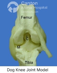
The cranial cruciate ligament (CCL) C is a key stabilizing structure of the stifle joint (equivalent to the human knee). It prevents excessive forward movement of the tibia (shin bone) relative to the femur (thigh bone), internal tibial rotation, and hyperextension of the stifle.
Two meniscus cartilages M within the joint act as cushions, help stabilize the knee and distribute nourishing joint fluid to the cartilage of the femur and tibia.
Surgical Treatment
"Surgery is generally recommended as soon as possible to minimize permanent joint damage and alleviate pain."
There are several surgical techniques for CCL repair, each with unique benefits and considerations. Your veterinarian will help determine the best option for your pet.
For additional surgical options, refer to "Cranial Cruciate Ligament Repair: Tibial Plateau Leveling Osteotomy (TPLO), Tibial Tuberosity Advancement (TTA), and TightRope® procedure."

How Modified Retinacular Imbrication Technique (MRIT) / Lateral Suture Stabilization (LSS) Works
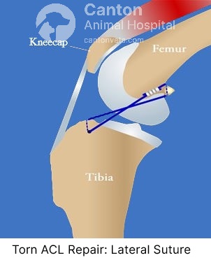
MRIT/LSS is an extracapsular stabilization technique, meaning the repair occurs outside the joint capsule rather than altering bone structure.
A strong synthetic suture, similar to medical-grade fishing line, is looped around the fabella (a small bone behind the femur) and secured through a drilled hole in the tibia.
This suture mimics the function of the original CCL, preventing abnormal forward movement of the tibia.
If the meniscus is torn or damaged, that part will be removed.
Over time, scar tissue forms, further stabilizing the joint and reducing reliance on the suture.
Expected Recovery Period
2 weeks post-surgery: Your pet should begin touching the toes to the ground while walking.
8 weeks post-surgery: Lameness should be mild to moderate.
6 months post-surgery: Your pet should remain near-normal limb function.
Postoperative Care
The limb may or may not be bandaged postoperatively, depending on the surgeon's preference.
Strict exercise restriction is required for the first 8 weeks, followed by a gradual return to normal activity.
The force exerted across the knee joint is four to five times body weight. Overuse can lead to implant failure before fibrous tissue develops.
If lameness does not improve within 8-12 weeks or worsens after initial improvement, there may be a torn meniscus or suture failure.
If necessary, imbricating sutures can be removed after 3 months without compromising stability, as fibrosis provides long-term joint support.
Prognosis
Approximately 85% of dogs show significant improvement post-surgery.
Around 50% regain normal limb function, while the other 50% may experience mild or intermittent lameness after intense activity.
While MRIT/LSS significantly improves limb function, it does not halt arthritis progression. Stiffness may occur in the morning, after exercise, or due to weather changes.
To manage joint stiffness and discomfort, veterinarians may recommend:
Chondroitin sulfate, MSM, and glucosamine supplements
Adequan injections (to promote joint health)
NSAIDs (Non-Steroidal Anti-Inflammatory Drugs) to reduce inflammation and pain.
At Canton Animal Hospital, we perform advanced orthopedic surgeries including TTA, TPLO, and Extracapsular Lateral Suture Stabilization (LSS) techniques. Each procedure is tailored to the individual needs of the patient, ensuring optimal recovery and long-term joint health
Extracapsular Lateral Suture (MRIT) – Best for small to medium-sized dogs.
Tibial Plateau Leveling Osteotomy (TPLO) – The gold standard for large, active dogs.
Tibial Tuberosity Advancement (TTA) – A less invasive alternative to TPLO.
Frequently Asked Questions (FAQs) about Lateral Suture Technique
Cranial Cruciate Ligament injuries and their treatment options, including the MRIT/LSS procedure, often raise questions among pet owners. Below are answers to some of the most commonly asked questions to help you understand the procedure, its benefits, and what to expect during recovery.
Related Articles about Surgeries
Is your pet scheduling surgery soon? Explore our resource center for additional articles that may be of interest.
Featured Resources

We Welcome New Patients!
We're always happy to give your furry friend care at our hospital. Get in touch today!
Contact UsHelpful Links
Additional Information


