Home | Surgical Info | Partial Rupture of Cranial Cruciate Ligament (CCL) in Dogs
Partial Rupture of Cranial Cruciate Ligament (CCL) in Dogs
Keywords:
Partial Rupture of Cranial Cruciate Ligament (CCL) in Dogs
A partial cranial cruciate ligament (CCL) tear is a common yet frequently overlooked cause of knee (stifle) lameness in dogs. While the ligament remains partially intact, it is still functionally compromised, leading to joint instability, pain, and an increased risk of arthritis. These injuries can be challenging to diagnose, as symptoms may mimic hip dysplasia or other orthopedic conditions.
At Canton Animal Hospital, we specialize in the early detection and treatment of partial CCL tears, ensuring your pet receives the best possible care before the injury worsens. Early intervention with advanced surgical techniques like TPLO or TTA can prevent a complete rupture and long-term joint damage.
Causes & Risk Factors
Common in young, large-breed dogs – Labrador Retrievers & Rottweilers are especially prone to partial CCL tears between 6 to 24 months of age.
Often bilateral – Many cases involve both knees, sometimes mimicking hip dysplasia.
Degenerative changes occur over time – Arthritis develops more slowly than in complete ruptures since the meniscus is often spared.
Two parts of the CCL – Partial tears may affect either:
Craniomedial Band (CrMB) – Taut in both flexion and extension.
Caudolateral Band (CdLB) – Taut only in extension.
Featured Resources

We Welcome New Patients!
We're always happy to give your furry friend care at our hospital. Get in touch today!
Contact Us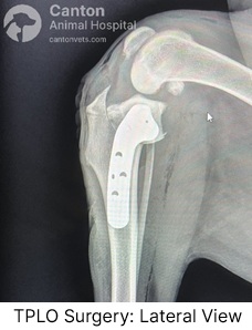
Diagnosis of Partial CCL Rupture
Signs & Tests Used for Diagnosis:
Mild to moderate lameness that mimics a complete tear but is less severe.
Pain during full knee extension, often with joint swelling (effusion).
Fat Pad Sign – A key radiographic finding indicating joint effusion.
Osteophyte Formation – Bone spurs indicating early arthritis.
MRI, ultrasound, or arthroscopy – Useful for detecting subtle ligament damage when physical exams are inconclusive.
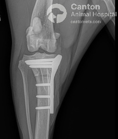
Treatment for Partial CCL Tears
Partial CCL tears should be treated like complete ruptures because the ligament is already functionally compromised.
Surgical Repair – TPLO (Tibial Plateau Leveling Osteotomy) or TTA (Tibial Tuberosity Advancement) are the gold standard for stabilizing the knee and preventing arthritis.
Early intervention is crucial – Delayed treatment increases the risk of meniscus damage and long-term joint degeneration.
Concerned about your dog’s knee health?
Schedule an orthopedic consultation at Canton Animal Hospital today!
Featured Resources

We Welcome New Patients!
We're always happy to give your furry friend care at our hospital. Get in touch today!
Contact UsTips and Advice from Our Team
Looking for advice about caring for your pet? Our blog features helpful tips and educational material from our team to support your needs.
