Home | Surgical Info | Cranial Cruciate Ligament Repair (Torn ACL Surgery) in Dogs
Cranial Cruciate Ligament Repair (Torn ACL Surgery) in Dogs
Keywords:
Cranial Cruciate Ligament (CCL) Repair in Dogs: Surgery & Treatment Options
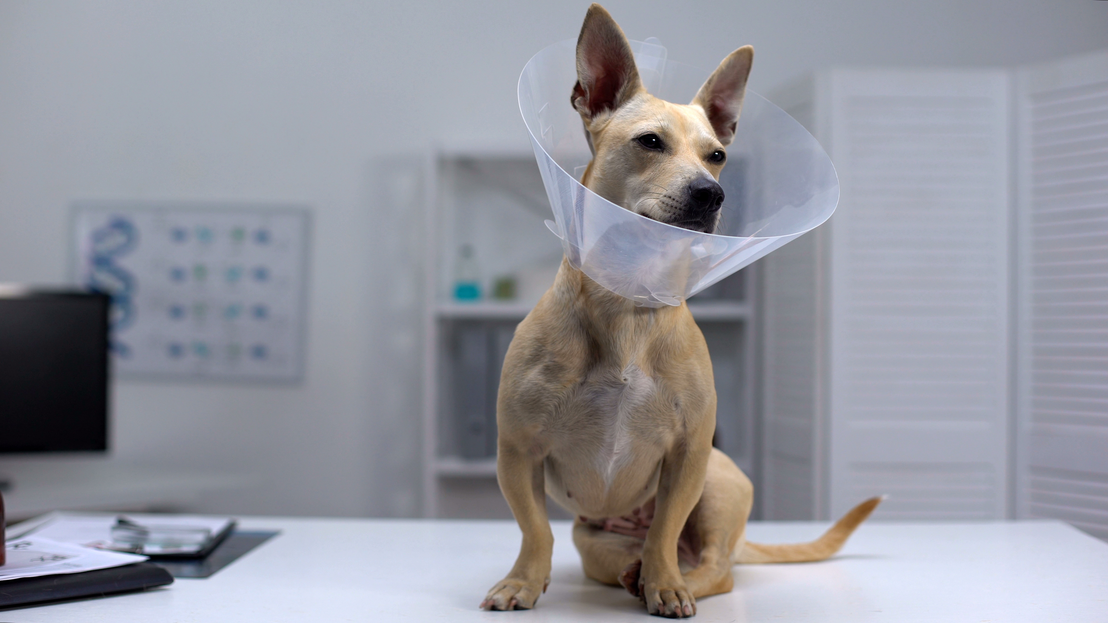
Cranial cruciate ligament (CCL) repair, also known as ACL surgery for dogs, is a common orthopedic procedure designed to stabilize the knee joint and restore mobility. A ruptured or torn CCL can lead to severe pain, lameness, and joint instability, ultimately causing osteoarthritis (OA) if left untreated.
Several surgical treatment options are available for CCL repair, including:
Extracapsular Lateral Suture (MRIT) – Best for small to medium-sized dogs.
Tibial Plateau Leveling Osteotomy (TPLO) – The gold standard for large, active dogs.
Tibial Tuberosity Advancement (TTA) – A less invasive alternative to TPLO.
Tightrope Technique – A minimally invasive option for specific cases.
Each procedure is tailored to the individual needs of the patient, ensuring optimal recovery and long-term joint health. Post-operative care plays a crucial role in the success of the surgery, helping to reduce pain, improve healing, and prevent further joint damage.
Is your dog struggling with a CCL injury? Contact Canton Animal Hospital today to explore the best treatment options!
What is the Cranial Cruciate Ligament (CCL)?
The Cranial Cruciate Ligament (CCL), similar to the ACL in humans, is a strong band of tissue that stabilizes the knee joint in dogs. It prevents excessive movement of the femur and tibia and ensures proper knee function. A torn or ruptured CCL leads to pain, joint instability, and arthritis if left untreated.
“In dogs, the most common knee injury is a rupture or tear of the cranial cruciate ligament. Most dogs with this injury cannot walk normally and experience pain. The resulting instability damages the cartilage and surrounding bones and leads to osteoarthritis (OA) in the knee joint. “
What Happens When a Dog Tears Its Cruciate Ligament?
Sudden pain and limping—Most dogs will avoid putting weight on the injured leg.
Intermittent use of the leg—Dogs may resume walking but continue to limp.
Joint instability—The tibia moves excessively, leading to cartilage damage and arthritis.
Meniscal damage—The C-shaped cartilage in the knee joint is prone to injury if left unstable.
Featured Resources

We Welcome New Patients!
We're always happy to give your furry friend care at our hospital. Get in touch today!
Contact Us

Why Do Cruciate Ligament Tears Occur?
Common Causes of ACL Ruptures in Dogs:
Trauma & Sudden Movement—Abrupt twisting, jumping, or missteps.
Degenerative Wear & Tear—Progressive ligament weakening due to genetics or age.
Obesity—Excess weight places added stress on the knee joint.
Breed Predisposition—Large breeds like Labradors, Rottweilers, and Newfoundlands are at higher risk.
Concurrent Joint Issues—Conditions like patellar luxation can contribute to ligament damage.
Why is Surgery Necessary for a Torn ACL?
A torn ACL in dogs does not heal on its own. Without surgery:
The knee remains unstable, leading to progressive joint damage.
Arthritis develops quickly, reducing mobility and causing chronic pain.
40-50% of dogs with one ACL tear will rupture the ligament in the other leg within two years.
Surgical intervention stabilizes the joint, reduces pain, and restores long-term mobility.
Common Surgical Techniques for ACL Repair in Dogs


What is the lateral suture stabilization (LSS/MRIT)?
The Lateral Suture Stabilization (LSS) or Modified Retinacular Imbrication Technique (MRIT) is a surgical procedure used to treat cranial cruciate ligament (CCL) injuries in dogs. It stabilizes the knee joint by placing a strong synthetic suture outside the joint to mimic the ligament’s function.
Uses a strong suture to mimic ligament function and stabilize the knee.
Best for small to medium-sized dogs.
Less invasive with a shorter recovery time.
Advantages of the LSS/MRIT Procedure
Less Invasive Surgery – No need to cut or reposition bones, leading to reduced surgical trauma.
Lower Cost – More affordable than TPLO or TTA procedures.
Effective for Small to Medium Dogs – Works well for dogs under 40 lbs. with moderate activity levels.
Faster Surgery & Recovery – Shorter procedure time with a recovery period of 8–12 weeks.
Fewer Implant-Related Complications
– No bone plates or screws, reducing the risk of implant failure.
Disadvantages of the LSS/MRIT Procedure
Less Effective for Large & Active Dogs – May not provide long-term stability for large breeds or highly active dogs.
Higher Risk of Suture Failure – Over time, the suture may stretch, loosen, or break, leading to joint instability.
Arthritis Progression – Does not alter knee biomechanics, so arthritis may still develop over time.
Long-Term Stability Concerns – Less durable than TPLO or TTA, especially for highly active dogs.
Rehabilitation Required – Strict post-op care and limited activity are necessary for a full recovery.


What is the TPLO procedure?
TPLO stands for tibial plateau leveling osteotomy. The Tibial Plateau Leveling Osteotomy (TPLO) is a widely used surgical technique to treat cranial cruciate ligament (CCL) injuries in dogs. It changes the knee’s biomechanics by altering the tibial slope, eliminating the need for the damaged ligament.
Gold standard for large-breed, active dogs.
Alters the tibial slope to eliminate instability without needing a new ligament.
Provides faster recovery and long-term success.
Advantages of the TPLO Procedure
Highly Effective for Long-Term Stability – Alters the knee’s biomechanics to restore function and reduce arthritis risk.
Best for Active & Large Breed Dogs – Provides strong, durable joint stability, ideal for working or athletic dogs.
Faster Return to Normal Activity – Many dogs start using the leg within days after surgery.
Lower Risk of Implant Failure – Well-studied with a high success rate in preventing future joint instability.
Reduces Arthritis Progression – Minimizes long-term joint deterioration compared to extracapsular repairs.
Disadvantages of the TPLO Procedure
Invasive Surgery – Requires cutting and repositioning the tibia, which may lead to more initial discomfort.
Longer Recovery Time – Full healing can take up to 12–16 weeks with strict post-op care.
Higher Cost – TPLO is more expensive than extracapsular repair techniques.
Potential for Complications – Risks include infections, bone fractures, implant loosening, or delayed healing.


What is the TTA procedure?
TTA stands for tibial tuberosity advancement. The procedure involves moving the patellar tendon attachment on the tibia forward which allows the quadriceps muscle to assume the normal job of the torn ACL by pulling forwards on the tibia to oppose the torn ACL.
Shifts the patellar tendon to stabilize the knee naturally.
Similar to TPLO but uses a different bone-cutting technique.
Reduces the risk of arthritis and promotes natural movement.
Advantages of the TTA Procedure
Faster Recovery – Many dogs bear weight within days, leading to quicker rehabilitation.
Less Postoperative Pain– May cause less discomfort compared to TPLO due to reduced bone cutting.
Improved Joint Stability – Alters knee biomechanics to minimize joint stress.
Lower Risk of Implant Failure – Some studies suggest fewer long-term implant complications than TPLO.
Ideal for Active & Large Dogs – Provides strong joint stability, making it suitable for athletic and working breeds.
Disadvantages of the TTA Procedure
Not Suitable for All Dogs – Dogs with extreme knee angles or small breeds may not be ideal candidates.
Risk of Bone Fractures – Cutting and shifting the tibial tuberosity can sometimes lead to fractures.
Implant-Related Complications – Metal implants can lead to infections, loosening, or irritation.
Post-Surgical Rehabilitation Required – A strict recovery plan is necessary for full healing.
Higher Cost – Generally more expensive than extracapsular repair techniques.


What Tightrope CCL Procedure for Torn ACL?
The Tight Rope Procedure is a variation of the lateral suture procedure.
Uses high-strength fiber implants to reinforce joint stability.
Less invasive alternative to TPLO and TTA.
Suitable for dogs with mild to moderate instability.
Advantages:
The suture material is anchored at more isometric points, allowing for a more natural range of motion while maintaining stifle stability.
Provides superior stabilization compared to traditional extracapsular techniques.
Minimally invasive compared to osteotomy-based techniques.
Disadvantages:
The use of a large, braided suture significantly increases the risk of serious infection.
Requires precise surgical technique and specialized equipment.
Post-Surgical Recovery & Rehabilitation
Proper post-operative care is essential for successful ACL repair:
Strict rest & limited activity for 6-12 weeks to allow healing.
Pain management with NSAIDs, nerve blocks, and CRI analgesia.
Physical therapy including passive range-of-motion exercises.
Weight management to reduce stress on the healing joint.
Cold & heat therapy to minimize swelling and enhance comfort.
Which ACL Surgery Is Best for My Dog?
There are multiple ACL repair techniques, and the best one depends on your dog’s size, activity level, and joint anatomy.
Surgery Type | Best For | Pros | Cons |
TPLO Surgery | Large & active dogs | Best long-term results | More expensive |
TTA Surgery | Medium to large dogs | Faster recovery | Not suitable for steep tibial angles |
Lateral Suture (MRIT) | Small & older dogs | Less invasive, lower cost | Higher failure rate in active dogs |
TightRope ACL Repair | Various breeds | Minimally invasive | Limited long-term studies |
Why Choose Canton Animal Hospital for ACL Surgery?
Affordable ACL Surgery—Providing high-quality orthopedic care at reasonable prices.
Expert Surgical Techniques—Offering TPLO, TTA, Tightrope, and Lateral Suture repairs.
State-of-the-Art Equipment—Advanced anesthesia monitoring, electrosurgical cautery, and precision orthopedic tools.
Pain Management Focused—Multi-modal strategies, including nerve blocks and post-op rehabilitation.
Open 7 Days a Week—Flexible scheduling and urgent orthopedic consultations available.
Featured Resources
We Welcome New Patients!
We're always happy to give your furry friend care at our hospital. Get in touch today!
Contact UsFrequently Asked Questions (FAQs)
Pet owners often have questions about ACL repair surgery for dogs. Here are answers to some of the most common concerns.
Tips and Advice From Our Team
Looking for advice about caring for your pet? Our blog features helpful tips and educational material from our team to support your needs.