Home | Surgeries | Hip Luxation in Dogs: Toggle Pin Repair
Hip Luxation in Dogs: Toggle Pin Repair
Hip Dislocation in Dogs
Hip luxation is a painful dislocation often caused by trauma. At Canton Animal Hospital, we offer Toggle Pin Repair, a surgical solution that restores joint stability and mobility. This procedure replaces the damaged ligament with a synthetic prosthesis, allowing immediate weight-bearing and reducing re-luxation risks to under 10%. With proper post-operative care, most dogs recover within 8-10 weeks. Contact Canton Animal Hospital in Michigan for expert evaluation and treatment.
Understanding Hip Luxation in Dogs:
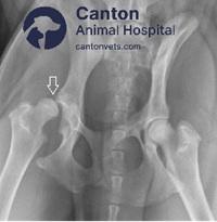
Coxo-femoral luxation (Hip Luxation) accounts for 90% of all luxations in dogs.
The most common cause of hip luxation is significant trauma, with most cases resulting from substantial injury.
Craniodorsal luxation is the most prevalent direction (80%) for coxo-femoral luxation, followed by ventral and then caudo-dorsal.
Featured Resources

We Welcome New Patients!
We're always happy to give your furry friend care at our hospital. Get in touch today!
Contact UsTreatment Options for Hip Luxation Include: ➜ Closed reduction ➜ Open reduction with toggle pin repair ➜ Femoral head and neck ostectomy (FHO) ➜ Total hip replacement (THR)
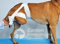
Closed reduction is recommended when the patient has minimal to no radiographic evidence of hip dysplasia and no concurrent orthopedic injuries. An Ehmer sling (or hobbles for ventral luxation) is advised for the first two weeks to prevent re-luxation, which occurs in 50% of closed reduction cases. Open reduction is indicated when closed reduction fails or if the patient has concurrent orthopedic injuries requiring immediate weight-bearing.
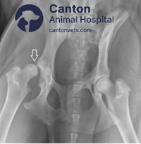
Several surgical methods are available for open reduction and stabilization of hip luxation.
The most widely used technique is the Toggle Pin (or Rod) Repair. This procedure replaces the ligament of the femoral head with a synthetic prosthesis, providing strong stabilization and allowing immediate weight-bearing without the need for an Ehmer sling. Long-term success rates are high, with reported re-luxation rates of less than 10% – the lowest among available treatments.
With any surgical stabilization of hip luxation, it is important to note that sutures and anchors serve as temporary solutions until the joint capsule and periarticular soft tissue heal. Patients with poor hip conformation are not ideal candidates for this method and should be considered for salvage procedures such as FHO or THR.
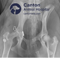
Surgical Procedure for Toggle Pin Repair
Craniolateral Approach to the Hip:
This procedure is performed using a craniolateral approach to the hip, with caudal retraction of the femur for complete visualization, exploration, and appropriate debridement of the acetabulum. After removing impinging tissue, clot, and the remaining round ligament, a hole is drilled through the medial wall of the acetabulum, centered in the acetabular fossa. A hand-held drill sleeve simplifies drilling while protecting the femoral head.
The diameter of the hole must be large enough for the chosen toggle pin and suture combination. A 3.2 mm toggle pin requires at least a 3.5 mm hole, but a 3.9 mm hole or larger is often necessary for heavy monofilament line. A 4.0 mm toggle pin typically requires a 4.5 mm to 5.5 mm drill bit. The toggle pin-suture combination should pass easily through the acetabular drill hole; if difficulty arises, enlarging the hole with a larger drill bit may be necessary.
Femoral Neck Tunnel for Suture
A tunnel is drilled from the subtrochanteric area of the lateral femur to the fovea capitis of the femoral head, or vice versa. The 3.5 mm drill bit is commonly used for medium to large dogs and is the minimum hole diameter used with the suture passer. Smaller patients require 2.0 mm or 2.7 mm tunnels.
Toggle Pin and Suture Insertion
The suture for repair is passed once through the toggle pin hole, creating a simple loop. The toggle pin is held with large needle holders, Kelly forceps, or a similar instrument. The suture is pulled tight along the sides of the toggle pin to seat within the grooves. The toggle pin is visually inserted into the acetabular drill hole as far as possible, then pushed completely through using the blunt end of a suture passer or drill bit.
Test Security of the Toggle Pin within the Pelvic Canal
The suture ends are spread and tensioned to pull the toggle pin tightly against the medial wall of the acetabulum. The toggle pin's secure seating within the pelvic canal is verified, and the suture is pulled through the femoral canal to exit the lateral femur while the femoral head is reduced into the acetabulum.
Verify Reduction and Secure the Suture
Appropriate reduction is confirmed, and the suture ends are tied over another toggle pin or a suture button. The hip should be well reduced and firmly seated without excessive tension on the suture, as this can compromise joint range of motion and lead to premature suture failure.
Convalescent Period
Your pet should begin touching toes to the ground at a walk within 10-14 days. Gradual resolution of lameness should follow. If your pet loses the ability to use the limb during recovery, please contact us immediately. Preventing slipping and excessive movement for the next 8 weeks is crucial. A sling is recommended for slippery surfaces, steps, or uneven ground.
Prognosis and Long-Term Success Rates
By 8-10 weeks post-surgery, the risk of re-luxation significantly decreases as the joint capsule heals. Your pet should regain normal mobility. Some discomfort may occur in cold weather or during weather changes, but long-term success rates are excellent, with re-luxation rates under 10%.
Post-Operative Care and Recovery
Administer prescribed antibiotics and pain medications as directed until completion.
If vomiting or inappetence occurs, discontinue medication and contact us.
Suture/staple removal should be performed in 10-14 days at our facility for progress evaluation.
Strict rest is required for 2 weeks. Excessive activity may lead to implant failure and unsuccessful hip healing. Keep your pet in a confined space until the next appointment.
Prevent running, jumping, slipping, falling, or playing.
Use a sling for support until staples/sutures are removed. Afterward, we will prescribe a rehabilitation program focused on leash walks.
Your pet’s health and your satisfaction are our top priorities. Please do not hesitate to call us with any concerns or questions.
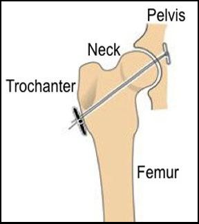
Toggle Pin Repair method involves placing a strand (or multiple strands) of suture material in a location that replicates the normal origin and insertion of the round ligament of the femoral head, which is torn when the hip is luxated.
The Toggle Pin Repair involves drilling two holes – one through the femoral head and neck from the third trochanter to the fovea capitis, and the other through the center of the acetabular fossa. The prosthetic ligament, often made of monofilament nylon or braided materials like LigaFiba Suture, is anchored to the medial wall of the acetabulum using a toggle pin and secured to the femoral neck with a knot, suture button, or second toggle pin.
Frequently Asked Questions: Toggle Pin Repair for Hip Luxation
Hip luxation can be overwhelming for both pets and their owners. Below are answers to common questions about toggle pin fixation surgery, recovery timelines, and what to expect post-op—so you can make confident, informed decisions about your dog’s orthopedic care.
Tips and Advice From Our Team
Our blog features helpful tips and educational material from our team to support your needs.
Featured Resources

We Welcome New Patients!
We're always happy to give your furry friend care at our hospital. Get in touch today!
Contact Us

