Home | Surgical Info | Cruciate Ligament Repair: MRIT Surgery
Cruciate Ligament Repair: MRIT Surgery
Mar 01 2022
Keywords:
What is MRIT surgery?
The MRIT procedure is short for Modified Retinacular Imbrication Technique, a surgical technique used for cruciate ligament repair in pets. During this procedure, an incision is made over the inside or outside aspect of the knee joint, and the joint is explored to assess the condition of the ligament and any associated injuries. If necessary, a meniscal injury may be partially removed. A sterile suture is then passed around the fabella and through a tunnel created in the tibia. The suture is tightened to stabilize the knee joint and prevent future injuries. This technique is known for its effectiveness in restoring stability and promoting a fast recovery in pets.
Cruciate ligament rupture is a common orthopedic condition that affects dogs and cats. The condition occurs when there is a partial or complete tear of the cranial cruciate ligament (CCL) in the stifle joint. CCL rupture can be caused by a traumatic injury or degenerative changes in the joint. Cruciate ligament repair is essential to restore the stability of the knee joint and prevent further damage. The MRIT technique is widely used to treat CCL injuries, and it has proven to be an effective surgical option in restoring stability and promoting fast recovery. The procedure has become increasingly popular over the years, amongst veterinarians due to its high success rate and low complication rate.
Featured Resources

We Welcome New Patients!
We're always happy to give your furry friend care at our hospital. Get in touch today!
Contact UsGeneral Information
a. Playing with other animals is not allowed during confinement. If there are other pets in your household, you will need to keep them separated. b. During confinement, your pet’s food intake needs to be reduced to help prevent weight gain. Most dogs will maintain their current weight if their food intake is cut in half. Water consumption should remain normal. c. The first two weeks following surgery you will need to monitor your pet’s incisions. Licking or chewing can cause infection or sutures to loosen. If you notice that your pet has started licking, you will need to take steps to discourage it from doing so. d. It takes a minimum of six to eight weeks for bones to heal. e. One of the most difficult aspects of confinement is that the animals will frequently feel better long before they are healed. At this point, your pet will start being more careless of the operated limb and is then more likely to be overactive and injure itself. Until the bone is healed, you must adhere strictly to the confinement guidelines and not allow your pet to do more. f. If your pet is jumping or bouncing in its confined area, it is being too active. Tranquilizers may be required to help alleviate your pet’s anxiety or control its activity. g. If at any time during your pet's recovery and healing it does anything that causes it to cry out or give a sharp yelp, contact your veterinarian. h. Following surgery your pet should always maintain its current level of function, or improve. If at any time during your pet’s recovery and healing it has a setback or decrease in function, contact your veterinarian. I. It is imperative that you inform your veterinarian at once if your pet does something that is potentially harmful to the surgery. If something has occurred that jeopardizes the outcome of surgery, it is usually less difficult to correct if it is caught right away, which leads to a better outcome for your pet. j. If your pet is too active during its confinement it may injure itself or slow healing which increases the amount of time your pet must be confined. k. Follow-up appointments are usually needed two weeks post-operatively to monitor incisions and healing. At eight weeks post-operatively radiographs are taken at which time your pet is started on a regulated activity regime. A final appointment at four months post-operatively is needed for additional radiographs and final instructions before returning your pet to normal activity.
Symptoms of Cranial Cruciate Ligament Disease
Limping
Holding the hind limb up
Sitting with the leg stuck out to the side
Stiffness, especially after exercise
Pain when the joint is moved or touched
Swelling of the joint
Sometimes clicking sound when walking
How is ACL Disease Diagnosed?
In order to diagnose ACL disease, a dog's medical history and a complete examination, including the "cranial drawer" and "tibial thrust" tests, are required to assess the integrity of the ACL. X-rays are also necessary to evaluate the level of arthritis and aid in determining which treatment is best for that pet. Sedation or anesthesia is necessary to make a definitive diagnosis and ensure the comfort of the pet.
Before undergoing cruciate surgery, dogs should have a correctly positioned x-ray to measure the slope of the tibia. This helps in making an informed decision about the appropriate type of surgery to perform. Based on our experience, TPLO surgery is a better option for dogs with a steep tibial slope, especially larger breeds. However, this may not be a significant factor in small breeds with a steep tibial slope. It is important for clients to be aware that a steep tibial slope puts greater force on the synthetic bands used in surgery (MRIT), which can lead to breakage.
Surgical Treatment: MRIT Procedure
There are several surgical techniques currently used to correct CCL rupture. You can learn more about each here: Tibial Plateau Leveling Osteotomy (TPLO), Tibial Tuberosity Advancement (TTA), Modified Retinacular Imbrication Technique (MRIT), and TightRope® procedure. Each procedure has unique advantages and potential drawbacks. Your veterinarian will guide you through the decision-making process and advise you on the best surgical option for your pet. Here we will focus specifically on the MRIT Procedure Technique.
Modified Retinacular Imbrication Technique (MRIT) / Lateral Suture Stabilization (LSS)
The MRIT /LSS technique is the oldest surgical correction for cruciate ligament injury in dogs. This technique is used mostly for small dogs and cats and has proven to show good outcomes in small-breed dogs. The name of the procedure originates from the fact that the joint is stabilized outside the joint capsule (externally).
CCL repair surgery typically consists of an initial examination of the inside of the knee. This examination may either be done by opening the joint capsule and looking inside or by using an arthroscope. Any damaged or torn portions of the CCL are removed. The shock absorber, or cartilage meniscus, that cushions the knee and sits between the femur and tibia, is examined. If the meniscus is torn or damaged, that part will be removed. After the joint capsule has been examined and any cartilage or ligament fragments are removed, the joint capsule is sutured closed.
“If the meniscus is torn or damaged, that part will be removed.”
In the MRIT procedure, a suture is passed from the outside/lateral aspect of the knee joint to the front of the tibia. Some surgeons refer to this as imbricating or overlapping, the extracapsular tissues to pull the joint tight and create stability to prevent front-to-back sliding of the femur and tibia. Usually, one or two bone channels or holes will be required to pass the suture from back to front. There have been several innovations in external capsular repair during the past decade. New materials, anchoring devices, and tools have allowed veterinary surgeons to perform this surgery more successfully than ever before. It is one of the extra-capsular techniques which means the function of the ACL, which is inside the joint, is replaced by placing a suture outside the joint. The suture most commonly a type of medical-grade "fishing line" is placed around the fabella and through the proximal tibia (Shin bone).
Expected Recovery Time
By 2 weeks after the surgery, your pet should be touching the toes to the ground at a walk. By 8 weeks, the lameness should be mild to moderate. By 6 months after the surgery, your pet should be using the limb well.
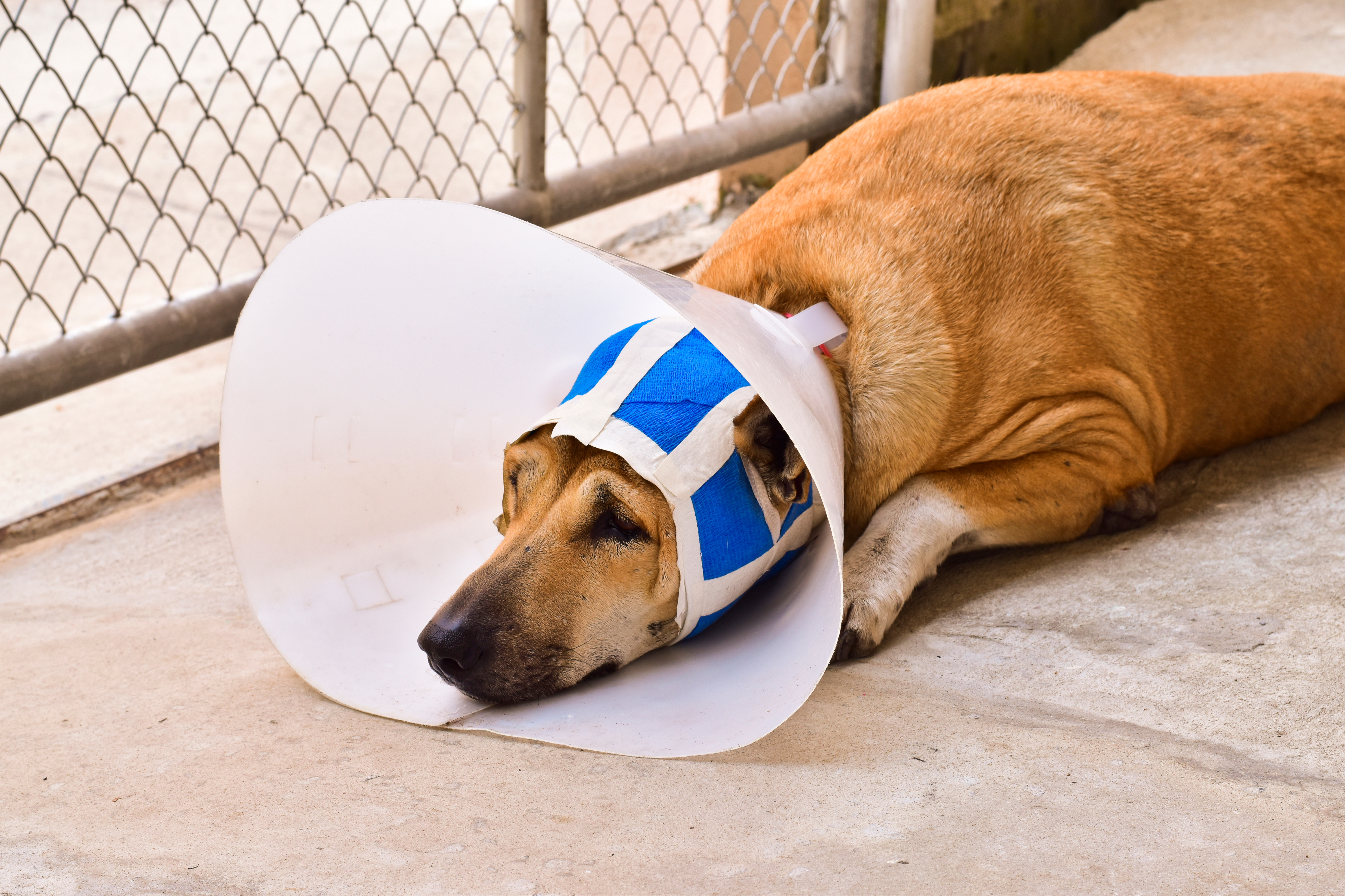
Post-Operative Care
After MRIT surgery, postoperative care is crucial for a successful recovery. The limb may or may not be bandaged postoperatively, depending on the surgeon's preference. To ensure proper healing, it's important to limit your pet's activity to very restricted exercise for the first 8 weeks. This will allow time for the bone to heal and the soft tissues to recover.
The muscle force across the knee joint is four to five times the body weight, and excessive activity may cause implant failure before the development of strong fibrous tissue. Therefore, it's important to follow our veterinarians' instructions carefully and not allow your pet to overexert themselves during the recovery period.
f your pet's lameness doesn't improve within 8 to 12 weeks after the surgery, or if it improves but then worsens, it may indicate a torn meniscus or a suture break or reaction. In this case, it's important to consult with our veterinary team for proper evaluation and treatment.
Prognosis
Extra-capsular techniques can significantly improve a dog's condition after a torn ACL, with around 85% of cases showing improvement from their preoperative state. However, it's important to note that with this technique, approximately 50% of dogs will still experience some degree of lameness, whether it's mild or intermittent following heavy activity. That said, around 50% of dogs will regain normal function of the limb. While extra-capsular techniques may not fully restore the limb to its pre-injury state, most dogs experience significant improvements in their condition compared to prior to surgery.
It's important to note that these techniques will not stop the progression of arthritis in the joint, so some stiffness may be present in the limb in the mornings, and your pet may experience some lameness after heavy exercise or during weather changes. Our veterinary clinic is dedicated to providing the highest quality care for your pet and will work with you to develop a comprehensive treatment plan tailored to your pet's individual needs.
After TTA surgery, some pets may experience stiffness. To help alleviate stiffness, our veterinary clinic may recommend the following options:
Chondroitin sulfate
MSM
glucosamine combinations
Adequan injections, and
NSAID (Non- Steroidal Analgesic Inflammatory Drug).
Our experienced veterinarians will carefully evaluate your pet's condition and recommend the best course of action. We will provide you with detailed instructions on how to administer any medications or supplements recommended for your pet's postoperative care.
Featured Resources
We Welcome New Patients!
We're always happy to give your furry friend care at our hospital. Get in touch today!
Contact UsTips and Advice From Our Team
Looking for advice about caring for your pet? Our blog features helpful tips and educational material from our team to support your needs.